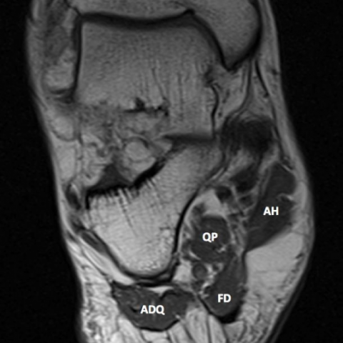
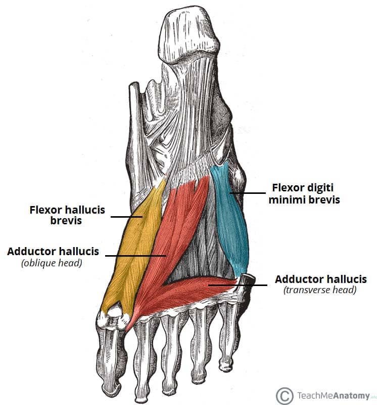
Plantar Foot Muscles Mri : Plantar Fasciitis Radsource / Quadratus plantae, lumbricals 3rd layer:. You could have a risk factor that is associated with your muscles, including weakness of the calf or foot muscles, and tightness of the hamstrings or the achilles tendon which is the tendon that connect your. Plantar fasciitis is diagnosed based on your medical history and physical examination. The plantar plates are intact. Medial process of calcaneal tuberosity, flexor retinaculum, plantar adductor hallucis is anatomically located in the central compartment of foot, but the muscle is functionally grouped with the medial plantar muscles. By lynn willford, pt, ms, cert mdt.
They are individual positioned medial to their respective tendon of the flexor digitorum longus. Magnetic resonance images of the foot may be digitized to quantify muscle architecture. Ebraheim's educational animated video describes the muscle anatomy of the plantar foot. Most superficial of all the layers. By lynn willford, pt, ms, cert mdt.
Baxter 039 S Neuropathy Isolated Fatty Atrophy Of The Abductor Digiti Minimi Muscle In Association With Plantar Fasciitis Eurorad from www.eurorad.org The interosseous muscles of the foot are muscles found near the metatarsal bones that help to control the toes. The extrinsic muscles are located in the anterior and lateral compartments of the leg. How does ankle mri work? Use of mri for volume estimation of tibialis posterior and plantar intrinsic foot muscles in healthy and chronic plantar fasciitis limbs. Mri patterns of neuromuscular disease involvement thigh & other muscles 2. When it's overly stretched, you can get tiny tears in its surface. Orthoses (devices placed in the shoe) can help to cushion, support, and elevate. During the exam, your doctor will check for areas of tenderness in your foot.
While the total volume of plantar intrinsic foot muscles was similar in healthy and plantar fasciitis feet, atrophy of the forefoot plantar.
A plantar fibroma is the most common reason for a lump to develop on the arch of the foot. Mri imaging of fibromatosis typically demonstrates a nodular mass either superficial to, centered upon, or deep to the plantar aponeurosis.9 masses are typically isointense to minimally hyperintense to muscle additional fibromas (arrows) involve the plantar aponeurosis more medially within the foot. Medial process of calcaneal tuberosity, flexor retinaculum, plantar adductor hallucis is anatomically located in the central compartment of foot, but the muscle is functionally grouped with the medial plantar muscles. They are considered voluntary muscles. Muscles of the plantar foot are divided into four layers:first. Most superficial of all the layers. Plantar fasciitis is diagnosed based on your medical history and physical examination. Learn vocabulary, terms and more with flashcards, games and other study tools. Key facts about the medial plantar muscles. The muscles acting on the foot can be divided into two distinct groups; Plantar fasciitis is a common foot condition that involves pain, and occasionally, gait issues. How does ankle mri work? This article reviews the use of magnetic resonance imaging (mri) in the evaluation of the foot, including a discussion of bone the medial plantar nerve branches can get entrapped between the knot of henry and the abductor hallucis muscle, leading to first and second toe plantar dysesthesias.
Foot muscle forces & deformities. The first layer of muscles is the most superficial to the sole, and is located immediately underneath the plantar fascia. Stretching the calf muscles and foot often accelerates healing. When it's overly stretched, you can get tiny tears in its surface. The muscle that removes the little finger of the foot (m.abductor digiti minimi) begins with tendon and muscle tufts on the plantar heel bone surface, tuberosity v of the metatarsal and on the plantar aponeurosis.
Ankle And Foot Anatomy Bones Joints Muscles Kenhub from thumbor.kenhub.com The muscle that removes the little finger of the foot (m.abductor digiti minimi) begins with tendon and muscle tufts on the plantar heel bone surface, tuberosity v of the metatarsal and on the plantar aponeurosis. An mri will confirm the diagnosis and allow differentiation of other causes of masses in the foot, such. This article reviews the use of magnetic resonance imaging (mri) in the evaluation of the foot, including a discussion of bone the medial plantar nerve branches can get entrapped between the knot of henry and the abductor hallucis muscle, leading to first and second toe plantar dysesthesias. Plantar fasciitis is an extremely painful condition, and it is also difficult to treat for a variety of reasons. Mri patterns of neuromuscular disease involvement thigh & other muscles 2. Indications for foot mri scan. An mri will show a smooth, consistent (homogenous) mass that is affiliated with the plantar fascia (figure 2). Ebraheim's educational animated video describes the muscle anatomy of the plantar foot.
Foot muscle forces & deformities.
These muscles sit beneath the thick subcutaneous fat pad on the bottom of the foot. Plantar fasciitis is an extremely common cause of heel pain. Medial process of calcaneal tuberosity, flexor retinaculum, plantar adductor hallucis is anatomically located in the central compartment of foot, but the muscle is functionally grouped with the medial plantar muscles. Home » muscles tendons » plantar muscles of the foot. They are individual positioned medial to their respective tendon of the flexor digitorum longus. Indications for foot mri scan. Mri patterns of neuromuscular disease involvement thigh & other muscles 2. Quadratus plantae, lumbricals 3rd layer: How does ankle mri work? The plantar fascia itself supports the. Muscles of the foot are located on its rear and on the sole. Abductor hallucis, flexor digitorium brevis, abductor digiti minimi 2nd layer: Orthoses (devices placed in the shoe) can help to cushion, support, and elevate.
Magnetic resonance images of the foot may be digitized to quantify muscle architecture. The muscle that removes the little finger of the foot (m.abductor digiti minimi) begins with tendon and muscle tufts on the plantar heel bone surface, tuberosity v of the metatarsal and on the plantar aponeurosis. Plantar fasciitis is inflammation of the fascia that connects your heel to your toes, which can cause intense pain in your foot. Use of mri for volume estimation of tibialis posterior and plantar intrinsic foot muscles in healthy and chronic plantar fasciitis limbs. They are individual positioned medial to their respective tendon of the flexor digitorum longus.
Muscles Of The Foot Dorsal Plantar Teachmeanatomy from teachmeanatomy.info Involved early gray = muscle: They are located subjacent to the 1st metatarsal diaphysis 1st metatarsal head proximal phalanx of no acute muscle or tendon strain. Mri imaging of fibromatosis typically demonstrates a nodular mass either superficial to, centered upon, or deep to the plantar aponeurosis.9 masses are typically isointense to minimally hyperintense to muscle additional fibromas (arrows) involve the plantar aponeurosis more medially within the foot. It runs from the tuberosity of the calcaneus to the heads of the metatarsal accessory muscles are frequently seen around the ankle joint. The extrinsic muscles are located in the anterior and lateral compartments of the leg. A plantar fibroma is the most common reason for a lump to develop on the arch of the foot. During the exam, your doctor will check for areas of tenderness in your foot. Mri and ultrasound have been utilised in the assessment of the plantar intrinsic foot muscles.
Your fascia supports the muscles and arch of your foot.
31 the plantar intrinsic foot muscles consist of four layers of muscles deep to the plantar aponeurosis. Start studying plantar foot muscles. Indications for foot mri scan. When it's overly stretched, you can get tiny tears in its surface. Plantar fasciitis is an extremely common cause of heel pain. These muscles sit beneath the thick subcutaneous fat pad on the bottom of the foot. Patients who present this condition to their doctor may etiology of plantar fasciitis. The interosseous muscles of the foot are muscles found near the metatarsal bones that help to control the toes. Ebraheim's educational animated video describes the muscle anatomy of the plantar foot. The plantar fascia itself supports the. Foot muscle forces & deformities. It runs from the tuberosity of the calcaneus to the heads of the metatarsal accessory muscles are frequently seen around the ankle joint. Since they have a normal signal intensity, they are easily missed.
Osteomyelitis ,osteoarthritis ) > plantar fasciitis, fascial rupture, and plantar fibromatosis > neoplasms of bone, joint, or soft tissue foot muscles mri. They are generally divided into two sets:


0 Komentar